
Radioembolization, commonly known as a Y90 procedure, is a minimally invasive treatment that combines embolization and radiation therapy to treat liver cancer. Embolization is a surgical technique that prevents blood flow to an area of the body to shrink a tumor or block an aneurysm. Radiation therapy is the use of a certain type of energy, called ionizing radiation, to kill cancer cells and shrink tumors. Tiny glass or resin beads filled with the radioactive isotope yttrium Y-90 are placed inside the blood vessels that feeds a tumor. This blocks the supply of blood to the cancer cells and delivers a high dose of radiation to the tumor while sparing normal tissue.
Radioembolization is used to treat tumors that were initially formed in the liver or have spread (or metastasized) to the liver from another part of the body. It is a palliative treatment, which means it does not provide a cure but instead helps slow down the growth of the disease and alleviate symptoms. The procedure is an option for patients who are not candidates for other treatments, including surgery or liver transplantation. It can help extend the lives of patients with inoperable tumors and significantly improve their quality of life.
How Does This Procedure Work?
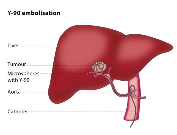
Using x-ray imaging and a contrast material to visualize the blood vessels, your radiologist inserts a catheter through the skin into a blood vessel and advances it to the treatment site. The radiation-filled microspheres, or glass beads, are then inserted through the catheter and advanced to the blood vessels supplying the tumor with blood.
Once the microspheres lodge at the tumor site, they deliver a high dosage of radiation directly to the cancer cells. The microspheres will block the flow of blood to the tumor, depriving the diseased cells of the oxygen and nutrients needed to grow.
There are two primary blood vessels that bring blood to the liver. Normal liver tissue receives about 75 percent of its blood supply from the portal vein and about 25% from the hepatic artery and its branches. When a tumor grows in the liver, it receives almost all of its blood supply from the hepatic artery. Because radioactive microspheres are delivered through the hepatic artery, they reach the tumor very directly while sparing most of the healthy liver tissue.
The radiation from yttrium-90 continually decreases over a two-week period and disappears after 30 days. The tiny microspheres remain in the liver without causing any problems.
How is the Procedure Performed?
This performed by a specially trained interventional radiologist in our interventional radiology suite.
An initial arteriogram is performed to visualize the upper abdominal arteries. At that time, arteries to areas of the stomach and duodenum which may have beads flow into them are closed with tiny coils of wire. At the end of the procedure, a bead with a nuclear medicine tracer on it is injected through the catheter to simulate the treatment. This will allow the interventional radiologist to calculate how much of the treatment dose can go to the lungs so that lung damage does not occur.
This procedure is often done on an outpatient basis. However, some patients may require admission following the procedure. This procedure is usually completed within an hour.
Talk with your doctor about any questions you may have for this procedure.