
At West Cancer Center & Research Institute, our Diagnostic Radiology Services provide a comprehensive range of imaging studies performed on-site for your convenience. Our state-of-the-art equipment ensures accurate, high-quality results. All images are interpreted by our in-house, sub-specialized radiologists, giving you expert insights from physicians who focus on specific areas of imaging. Our collaborative approach helps your care team make precise diagnoses and develop personalized treatment plans—all in one location.
Appointment Information:
To refer a patient or schedule an appointment, please contact our Radiology Department directly using the information below, or call us at 901.609.3556. Our team is ready to assist you with any questions and ensure a seamless scheduling experience.
CARDIAC CALCIUM SCORE
HOW IT WORKS: A CT scan measures calcium deposits in the coronary arteries, which can indicate plaque buildup.
WHAT IT IS USED FOR: It assesses your risk for heart disease and helps guide preventive care.
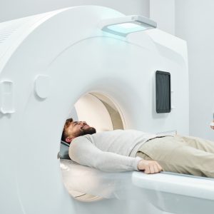
WHAT TO EXPECT: You’ll lie on a table while the scanner takes images of your heart. No injections or contrast are needed, and the test is quick—about 10 minutes.
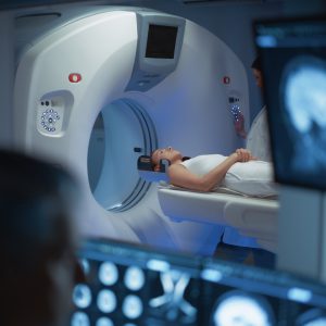
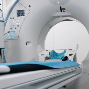
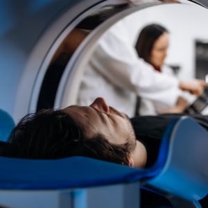
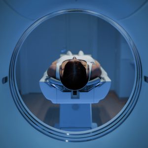
CT (COMPUTED TOMOGRAPHY)
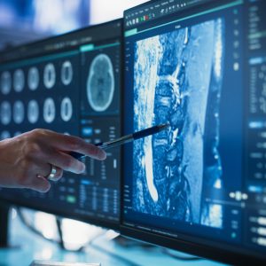
HOW IT WORKS: CT uses X-rays and computer processing to create cross-sectional images of the body.
WHAT IT IS USED FOR: It helps diagnose conditions like infections, tumors, and injuries.
WHAT TO EXPECT: You’ll lie on a table that moves through a donut-shaped scanner. Sometimes contrast dye is used. The scan is painless and usually takes 10–30 minutes.
FULL BODY MRI
HOW IT WORKS: MRI uses magnetic fields and radio waves to create detailed images without radiation.
WHAT IT IS USED FOR: It screens for tumors, inflammation, or other abnormalities throughout the body.
WHAT TO EXPECT: You’ll lie in a tube-like scanner for 45–90 minutes. It’s painless but noisy; ear protection is provided.
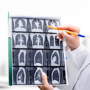
LOW DOSE CT LUNG SCREENING
HOW IT WORKS: A CT scan with lower radiation dose images the lungs.
WHAT IT IS USED FOR: It detects early lung cancer in high-risk patients (like smokers).
WHAT TO EXPECT: You’ll lie on a table for a quick scan—usually under 10 minutes.
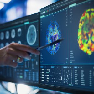
MRI
HOW IT WORKS: Uses magnets and radio waves to produce detailed images of organs and tissues.
WHAT IT IS USED FOR: Common for brain, spine, joints, and soft tissue evaluation.
WHAT TO EXPECT: Similar to full-body MRI but focused on one area. Duration: 30–60 minutes.
NETSPOT SCAN
HOW IT WORKS: Uses a radioactive tracer (Gallium-68) for PET imaging to detect neuroendocrine tumors.
WHAT IT IS USED FOR: Locates and stages neuroendocrine tumors.
WHAT TO EXPECT: Injection of tracer, wait about an hour, then PET scan for 30–60 minutes.
NUCLEAR MEDICINE BONE SCAN
HOW IT WORKS: A small amount of radioactive tracer is injected and absorbed by bones. A special camera detects areas of abnormal activity.
WHAT IT IS USED FOR: It checks for bone damage, cancer spread, or infection.
WHAT TO EXPECT: After the injection, you’ll wait 2–4 hours before scanning. The scan itself takes about 30–60 minutes.
PET (POSITRON EMISSION TOMOGRAPHY) SCAN
HOW IT WORKS: Uses a radioactive sugar tracer to show metabolic activity in tissues.
WHAT IT IS USED FOR: Detects cancer, monitors treatment, and evaluates brain or heart function.
WHAT TO EXPECT: Injection, waiting period, then scan lasting 30–60 minutes.
PSMA PET IMAGING
HOW IT WORKS: PSMA PET uses a small amount of radioactive tracer that binds to prostate-specific membrane antigen (PSMA) on prostate cancer cells. A PET scanner detects the tracer to create detailed images.
WHAT IT IS USED FOR: It helps locate prostate cancer cells in the body, especially for staging or detecting recurrence.
WHAT TO EXPECT: You’ll receive an injection of the tracer, wait about an hour, then lie still on a table during the scan. The process is painless and usually takes 30–60 minutes.

ULTRASOUND
HOW IT WORKS: Uses sound waves to create images of organs and tissues.
WHAT IT IS USED FOR: Evaluates organs, blood flow, and guides procedures.
WHAT TO EXPECT: A gel is applied to the skin, and a handheld probe moves over the area. Painless and usually 15–30 minutes.
X-RAY
HOW IT WORKS: Uses a small dose of radiation to create images of bones and some organs.
WHAT IT IS USED FOR: Checks for fractures, infections, or lung conditions.
WHAT TO EXPECT: Quick and painless — usually takes just a few minutes.