
A DEXA scan is a high-precision type of X-ray that measures your bone mineral density and bone loss. If your bone density is lower than normal for your age, it indicates a risk for osteoporosis and bone fractures.
What is a DEXA Scan?
DEXA stands for Dual Energy X-ray Absorptiometry. This technique was introduced for commercial use in 1987. It sends two X-ray beams at different peak energy frequencies to the target bones.
One peak is absorbed by soft tissue and the other by bone. When the soft tissue absorption amount is subtracted from the total absorption, the remainder is your bone mineral density. Standard X-ray diagnostics used before the development of the DEXA technology were only able to detect bone loss that was greater than 40 percent. DEXA can measure within 2 percent to 4 percent precision.
The test is noninvasive, fast, and more accurate than a regular X-ray. It also involves an extremely low level of radiation.
The World Health Organization (WHO) established DEXA as the best technique for assessing bone mineral density in postmenopausal women. DEXA is also known as DXA or bone densitometry.
How to prepare for your DEXA scan?
DEXA scans are usually outpatient procedures. There aren’t any special preparations needed, except to stop taking any calcium supplements for 24 hours before the test.
Wear comfortable clothing. Depending on the body area being scanned, you may have to take off any clothes with metal fasteners, zippers, or hooks. The technician may ask you to remove any jewelry or other items, such as keys, that may contain metal. You may be given a hospital gown to wear during the exam.
Let your doctor know in advance if you’ve had a CT scan requiring use of a contrast material or had a barium exam. They may ask you to wait a few days before scheduling a DEXA scan.
You should let the doctor know if you’re pregnant or suspect you might be pregnant. They may want to defer the DEXA scan until after you have the baby or take special precautions.
What to Expect:
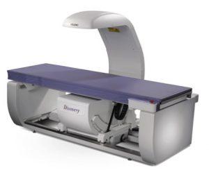
The DEXA apparatus includes a flat padded table that you lie on. A movable arm above holds the X-ray detector. A device that produces X-rays is below the table.
The technician will position you on the table. They may place a wedge under your knees to help flatten your spine for the image, or to position your hip. They may also position your arm for scanning.
The technician will ask you to hold very still while the imaging arm above slowly moves across your body. The X-ray radiation level is low enough to allow the technician to remain in the room with you while operating the device.
The whole process takes just a few minutes.
Your Results:
Your DEXA results will be read by a radiologist and given to you and your doctor in a few days.
The scoring system for the scan measures your bone loss against that of a healthy young adult, according to standards established by the WHO. This is called your T score. It’s the standard deviation between your measured bone loss and the average.
- A score of -1 or above is considered normal.
- A score between -1.1 and -2.4 is considered as osteopenia, increased risk for fracture.
- A score of -2.5 and below is considered as osteoporosis, high risk for fracture.
Your results may also give you a Z score, which compares your bone loss to that of others in your age group.
The T score is a measure of relative risk, not a prediction that you’ll have a fracture.
Your doctor will go over the tests results with you. They’ll discuss whether treatment is necessary, and what your treatment options are.